3-Microtomography
PRESENTATION
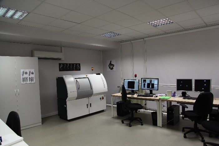
X-ray microtomography is an imaging technique allowing the 3D reconstruction of a sample. The method consists in reconstructing a volume from 360° X-rays. A large number of X-rays of the rotating object are thus taken and reflect a value linked to the absorption coefficient of the material, at each position in space. The AniRA-ImmOs platform has available a Phoenix Nanotom S Microtomograph.
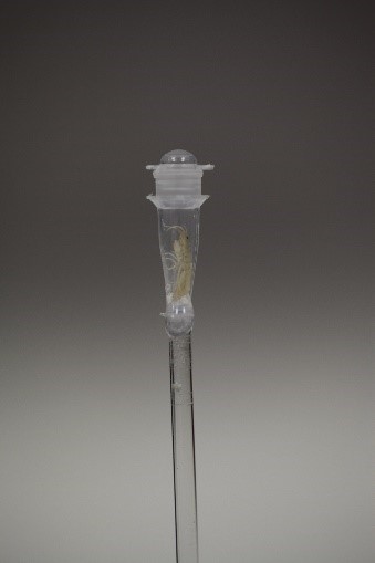
This instrument allows the acquisition of three-dimensional data with very high resolution, reaching micrometres, for samples size in the millimetre range.
Possible applications:
- NON-DESTRUCTIVE STUDIES OF THE INTERNAL STRUCTURE OF AN OBJECT
- Bone imaging and extraction of internal structures of interest
- Soft tissue imaging (lungs, plants, etc.)
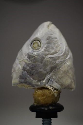
- Imaging of fossilised samples
- MORPHOMETRIC GEOMETRY
- Comparative morphological analyses. Micro-architecture parameters: BMD, BV / TV, cortical, trabecular….
- 3D reconstruction of fossils, etc.
ACTIVITIES
We offer training for X-rays images acquisitions and 3D reconstruction of the sample. We provide advice on acquisition parameters optimisation.
EQUIPEMENTS
The platform is equipped with a Phoenix Nanotom S X-Ray Computed Tomograph
Technical characteristics:
Imaging
The resolution of the equipment is around 1 μm. The minimum voxel size is < 0.5 μm. The equipment allows for edge enhancement by using phase contrast. The maximum field of view is 60 mm by 60 mm (for virtual enlargement of the field of view see 'Detector').
X-ray generator
The equipment includes a high power nanofocus X-ray tube xs|180nf with a maximum output power of 15 W and a maximum high voltage of 180 kV. The minimum distance between focus and sample is 0.4 mm.
X-ray detector
The 2D detector consists of 2300 x 2300 pixels and has an active area of 120 mm by 120 mm (corresponding to a pixel size of 50 μm x 50 μm). The dynamic range is 850:1.
The detector can be moved in horizontal direction for a virtual enlargement of the detector (travel range ca. 240 mm which corresponds to a virtual detector size of 360 mm by 120 mm)
CONTACT / LOCATION
For any information, please contact us:
S. Djebali : immos-sfrbiosciences.lyon@inserm.fr
Location of the instrument: ENS de Lyon (monod site)
PRATICAL INFORMATIONS
All types of samples can be analysed as long as their density allows sufficient attenuation of X-ray photon radiation.
- The microtomograph is accessible for half-day sessions (8 a.m.-1 p.m. or 1 p.m.-6 p.m.).
- For new users: Please contact the facility staff ( immos-sfrbiosciences.lyon@inserm.fr ). We will guide you to find the most suitable technology.
- Training: two training courses are mandatory before accessing the booking system and independently use the microtomograph. The courses include one theoretical course on awareness of the risks associated to X-rays and one practical course to learn about the equipment (approximately 1 day).
- Booking site: The equipment can be booked online upon training : https://ifr128.univ-lyon1.fr/resas/immos/select.php
 |
 |
 |
 |


